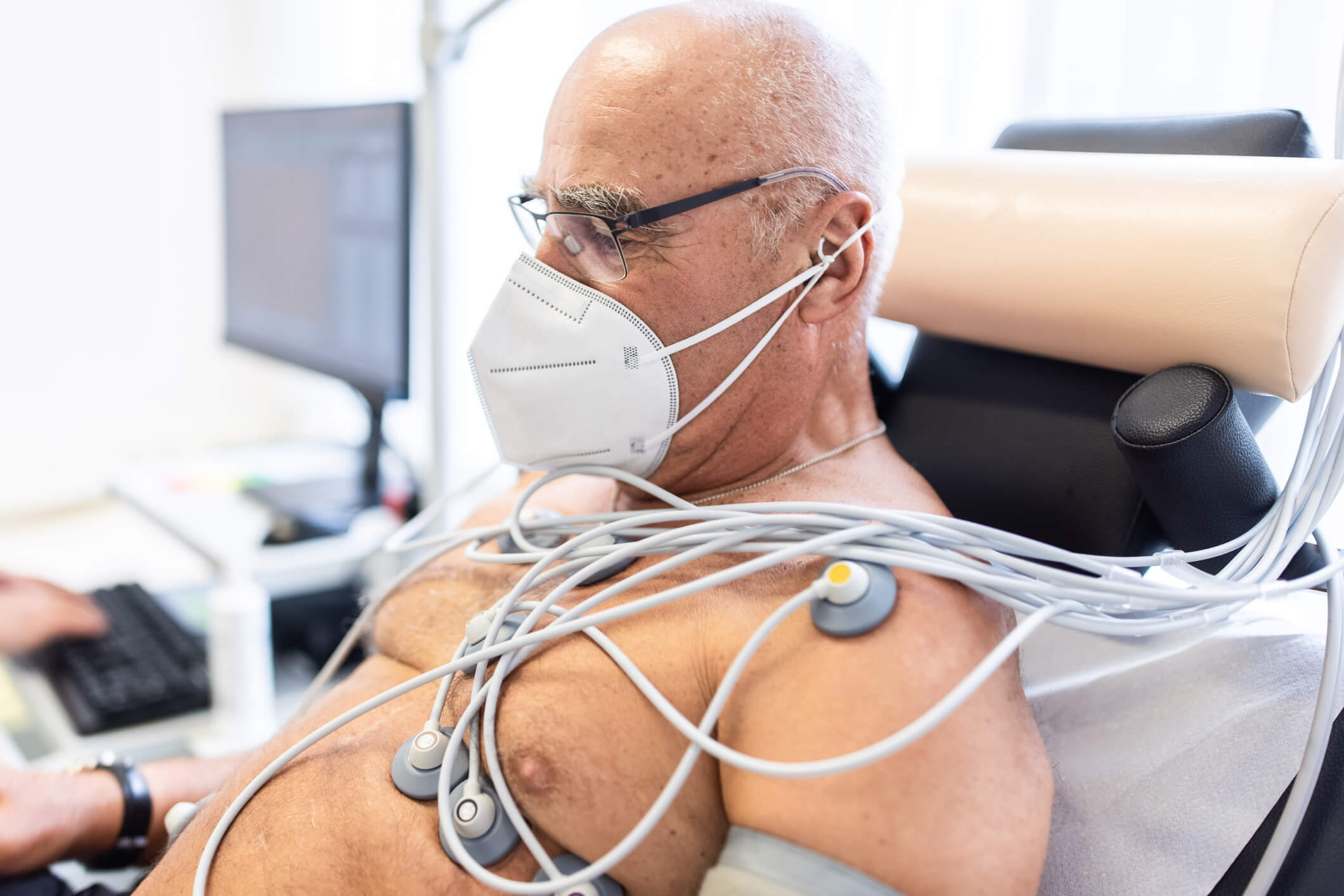
SARS-CoV-2, the virus that causes COVID-19 infection, damages the lungs, impairing gas exchange and depriving the body of oxygen. It also has significant cardiovascular effects, particularly in patients with preexisting cardiovascular disease (CVD). The reduced oxygen supply to the heart muscle causes inflammation, tissue damage, and arrhythmias and decreases cardiac output. Infection and inflammation further impair cardiac biochemical and fibrinogen pathways and diminish cardiac muscle integrity, leading to myocardial injury and dysfunction while also increasing the metabolic demand on many organs, including the heart.
Heart damage caused by COVID-19 without other causes is referred to as myocardial infarction type 2.1 It is diagnosed using cardiac biomarker measurements, such as high-sensitivity cardiac troponin l (cTnI) and N-terminal pro-B-type natriuretic peptide (NT-proBNP). Treatment is based on increasing knowledge of the SARS-CoV-2 life cycle and its association with host cells and involves preventing exposure, diminishing viral proliferation, and attenuating inflammatory responses. Educating patients on available treatment options, including potential side effects and complications, is important.
Cardiovascular Complications of COVID-19
COVID-19 induces a range of negative cardiac effects, such as hypoxic respiratory failure and hypoxemia leading to hypoxia-induced myocardial injury (3%–33%), microvascular injury, venous thromboembolism (23%–27%), causing small vessel ischemia, and pulmonary embolism, which can result in acute right ventricular failure [33%–47%], and left ventricular dysfunction (10%–41%).2 Other cardiac complications of COVID-19 include arrhythmias (9%–17%), (eg, atrial and ventricular fibrillation), ventricular tachyarrhythmia, myocarditis, acute myocardial injury, and venous thromboembolism.
Preexisting CVD increases the risk for these complications. In a New York City area analysis of 5700 patients hospitalized with COVID-19, the most common comorbidities were hypertension (57%), obesity (42%), and diabetes (34%).3 In a meta-analysis of 6 studies on cardiometabolic comorbidities of COVID-19 in China (N=1527;), the most common cooccurring diseases were hypertension (17.1%), cardia-cerebrovascular disease (16.4%), and diabetes (9.7%).4 Cardiovascular diseases — hypertension, in particular — are also associated with the highest morbidity rate (10.5%) after the development of COVID-19.3 Other comorbidities, such as chronic pulmonary disease, diabetes, and cancer, increase mortality as well.
Regardless of the presence of preexisting conditions, severe inflammation of the heart muscle can cause life-threatening myocarditis.1 Case studies show that COVID-19 viral infection injures cardiomyocytes, leading to acute myocardial injury in 8% patients.4 Acute cardiac injury, as shown by elevated cTnI levels, occurs in 8% to 62% of patients hospitalized with COVID-19 and is associated with greater severity, the need for mechanical ventilation, and death.2 In Wuhan, China, 5 of the first 41 patients diagnosed with COVID-19 had elevated cTnI levels (>28 pg/mL).3
Hospitalized patients with COVID-19 frequently present with acute cardiac compromise, as demonstrated by acute heart failure (3%-33%), ventricular dysfunction (right, 33%-47%; left, 10%-41%; biventricular, 3%-15%), venous thromboembolism (23%-27%), cardiogenic shock (9%-17%), arrhythmias (9%-17%), myocardial ischemia or infarction (0.9%-11%), stress cardiomyopathy (2%-5.6%), and arterial thrombosis secondary to viral-mediated coagulopathy.2 In a recent report on 138 individuals hospitalized with COVID-19, arrhythmias were the second most common serious complication after acute respiratory distress syndrome, occurring in 16.7% of patients.3 Arrhythmias were present in 44% of patients who required treatment in the intensive care unit (ICU) and 7% of those who did not.3
Even in the absence of COVID-19-related lung damage and after the acute phase of illness has resolved, healthy adults can experience cardiac problems.1 Such extrapulmonary manifestations can have long-term consequences.2
Pathophysiology
The inflammatory response to infection interferes with cardiac biochemical pathways, diminishes the integrity of cardiac muscle, and initiates abnormal clotting cascades, leading to myocardial injury and dysfunction.
SARS-CoV-2 enters host cells via a viral spike protein and angiotensin-converting enzyme 2 (ACE2) receptors.1 ACE2 has broad expression in the heart, lungs, gastrointestinal system, and kidneys and is vital in the neurohumoral management of the cardiovascular system. It regulates the renin-angiotensin-aldosterone system, which converts angiotensin II, a vasoconstrictor and proinflammatory mediator that can damage the capillary endothelium, to angiotensin, a vasodilator. As the virus binds with and down-regulates ACE2 to infiltrate cardiac myocytes and alveolar epithelial cells, levels of angiotensin II increase, causing lung and heart injury.1
The inflammatory response disrupts the coagulation cascade, leading to coagulopathy, disseminated intravascular coagulation (DIC), and the formation of pulmonary and cardiovascular emboli.5 Initially, this is evidenced by elevated levels of D-dimer and fibrinogen/fibrin degradation products and abnormalities in coagulopathy parameters. In DIC, micro-clots form in the blood and can become lodged in the lungs, arteries, other blood vessels, or extremities, leading to complications. DIC also interferes with the body’s ability to dissolve the clots and depletes platelets and clotting factors, presenting challenges in patients with bleeding problems.
Diagnosis
Cardiac injury is measured by cTnl and NT-proBNP levels. The significant relationship between these biomarkers demonstrates the close association of inflammation and myocardial stress. Elevated cTnl is a predictive marker for COVID-19, regardless of the presence of pre-existing CVD. Myocardial injury also confirms the presence of more severe systemic inflammation, further demonstrated by increased levels of other biomarkers, such as leukocyte counts, procalcitonin, creatine kinase, and myoglobin.1
Cardiac injury is strongly associated with worse COVID-19 outcomes, as evidenced by a trend toward rising serial cTnl and NT-proBNP measurements in individuals with poor clinical outcomes vs those who recover. A recent review demonstrated a significant difference in in-hospital mortality rates for patients who had increased cTnI levels compared with those who did not (51.2% vs 4.5%, respectively).1 Another review showed elevated cTnl levels in few survivors of uncomplicated COVID-19 (1%-20%), many severely ill patients (46%-100%), and nearly all ICU-level patients and non-survivors.2 Autopsies have revealed evidence of COVID-19-associated lymphocytic myocarditis (14%-40%), , small vessel thrombosis (19%), focal pericarditis (19%), endocardial thrombosis (14%), and right ventricular strain (19%).2
COVID-19 is responsible for progressive systemic inflammation leading to respiratory distress, sepsis, multiorgan failure, and death. Studies to date demonstrate a delay between symptom onset and myocardial damage; therefore, cardiac magnetic resonance imaging (MRI) is used to detect typical signals of acute myocardial injury. The gold standard diagnostic test, endomyocardial biopsy (EMB), identifies myocyte necrosis and mononuclear cell infiltrates with viral causes; myocarditis can also have other autoimmune-mediated causes. Biopsy studies in European patients with acute myocarditis, for example, found viral etiology in 37.8% to 77.4% of cases. Evidence describing myocardial injury in COVID-19 is scarce and based on individual case studies, highlighting the need for systematic assessment.3
Treatment Options
Treatments for COVID-19 have been developed, but the illness remains incurable.1 Preventive and therapeutic strategies are based on the sequence of pathogenic events and increasing understanding of the SARS-CoV-2 life cycle. The overarching goal is the prevention of COVID-19 via social distancing, mask wearing, and vaccination. Recent randomized trials for the mRNA vaccine have “reported ≈95% efficacy with very low incidence of serious adverse events demonstrated across race, ethnicity, and age groups.”2 However, long-term safety and durability, as well as efficiency in different populations, remain key research topics.
The next steps in treatment are inhibiting viral proliferation and impeding inflammatory responses in the body. This involves 3 subclasses of therapies: repurposed previously approved therapeutics, biologics, and small-molecule therapeutics. Prevention of virus–host cell receptor binding is a common approach to decreasing virus proliferation and involves targeting the COVID-19 spike protein or ACE2. This has been achieved using a lipopeptide to negate cell–cell fusion via the spike protein, to disrupt the spread of the virus into airway epithelial cells.2 Other entry inhibitors have been repurposed from existing therapeutics, such as clemastine, trimeprazine, amiodarone, bosutinib, flupenthixol, toremifene, and azelastine.2 In emergent situations, antiviral treatment with remdesivir, nirmatrelvir-ritonavir, or molnupiravir may diminish the severity of COVID-19 complications, but these medications must be administered soon after symptom onset to be effective: 7 days for remdesivir and 5 days for nirmatrelvir-ritonavir and molnupiravir.6 In addition, steroids, hydroxychloroquine, i.v. immunoglobins, and active mechanical life support have been used to treat COVID-19.
Finally, organ-specific therapies are used to improve complications, such as a prothrombotic state, acute kidney injury, or stroke syndromes. Continuous heparin infusion along with additional anticoagulation therapy can inhibit microvascular injury and thrombosis, preventing small-vessel ischemia, pulmonary embolism, or stroke syndromes. Continuous renal replacement therapy filters blood and removes waste, allowing the kidneys to recover from acute injury. Extracorporeal membrane oxygenation provides cardiac and respiratory support when the lungs are unable to provide adequate gas exchange or perfusion. Even with these advanced processes and devices, outcomes are bleak for those requiring such strategies.
Education
The long-term cardiac consequences of COVID-19-associated myocardial injury include evidence of myocardial fibrosis or myocarditis in 9% to 78% of patients after acute COVID-19 infection.2 Although evidence-based recommendations for post-COVID-19 follow-up evaluations are lacking, patients with cardiac involvement should receive close monitoring every 1 to 3 months, including 12-lead and Doppler echocardiograms.3 Continued monitoring and cardiac plans, including medication regimens, scheduled follow-up visits, referrals, and upcoming procedures, should be in place for any patient with cardiac complications of COVID-19. Patients should receive education about this to support adherence.
Health care providers also require ongoing education on the appropriate management of COVID-19 and its complications. For example, some physicians prescribe the antibiotic azithromycin to decrease the severity of the virus. Unfortunately, azithromycin is known to cause QT prolongation, which can lead to torsades de pointe and serious arrhythmias and increase the risk for sudden death. The use of other medications, including chloroquine and hydroxychloroquine, has been suggested; however not enough clinical data exist to support this approach. If these agents are prescribed, recipients should be monitored for QT prolongation and other adverse effects.1
Limitations
Because of the lack of long-term research on COVID-19, current studies may be limited by inclusion bias or small cohorts. Information based on early population exposure and untested medications and methods has likely evolved. Other factors affecting study applicability include the finite nature of hospital resources, including staffing, and uneven access to testing kits and treatment, which initially limited the categories of people tested. In addition, asymptomatic individuals who have been inoculated may not seek testing or treatment. These realities contribute to the challenge of assessing the true occurrence, prevalence, and mortality of COVID-19.3
Conclusion
The mechanisms underlying COVID-19-related myocardial injury remain unclear, with questions around systemic vs local reactions and the initiation of ischemic vs inflammatory processes yet to be answered. However, clinical evidence indicates that COVID-19 negatively affects the cardiac system. Similarly, other acute viral infections are well documented to cause cardiac injury and acute myocarditis.3 Preexisting cardiac diseases such as hypertension and CVD increase the risk for COVID-19-related complications; and comorbidities such as obesity, pulmonary diseases, and diabetes increase the complexity of the illness and are associated with worse clinical outcomes.
Treatment involves prevention of the virus via vaccination and social distancing and medications to decrease the absorption and spread of COVID-19 throughout the body. Education on the effects of current medication therapies and monitoring of cardiac function after recovery are important for the long-term management of this illness.
Seva McKee, RN, BSN, practices in the cardiac catheterization laboratory and is working on her doctor of nursing practice degree at the University of North Florida in Jacksonville.
References
1. Soumya RS, Unni TG, Raghu KG. Impact of COVID-19 on the cardiovascular system: a review of available reports. Cardiovasc Drugs Ther. Published online September 14, 2020. doi: 10.1007/s10557-020-07073-y
2. Chung MK, Zidar DA, Bristow MR, et al. COVID-19 and cardiovascular disease: from bench to bedside. Circ Res. Published online April 16, 2021. doi: 10.1161/CIRCRESAHA.121.317997
3. Guzik TJ, Mohiddin SA, Dimarco A, et. al. COVID-19 and the cardiovascular system: implications for risk assessment, diagnosis, and treatment options. Cardiovasc Res. Published online April 30, 2020. doi: 10.1093/cvr/cvaa106
4. Li B, Yang J, Zhao F, et al. Prevalence and impact of cardiovascular metabolic diseases on COVID-19 in China. Clin Res Cardiol. Published online March 11, 2020. doi:10.1007/s00392-020-01626-9
5. Srivastava S, Garg I, Bansal A, Kumar B. COVID-19 infection and thrombosis. Clin Chim Acta. Published online July 24, 2020. doi: 10.1016/j.cca.2020.07.046
6. Centers for Disease Control and Prevention. COVID-19 treatments and medications. Updated May 26, 2023. Accessed July 19, 2023. https://www.cdc.gov/coronavirus/2019-ncov/your-health/treatments-for-severe-illness.html
This article originally appeared on Clinical Advisor
